 京公网安备11010802034965号
京ICP备13020181号-2
京公网安备11010802034965号
京ICP备13020181号-2
《中国塑料》编辑部 ©2008-2024 版权所有
地址:北京市海淀区阜成路11号 邮编:100048
编辑部:010-68985541 联系信箱:cp@plaschina.com.cn
广告部/发行部:010-68985253 本系统由北京玛格泰克科技发展有限公司设计开发

中国塑料 ›› 2021, Vol. 35 ›› Issue (9): 136-146.DOI: 10.19491/j.issn.1001-9278.2021.09.021
韦宗辰1, 郗悦玮1,2( ), 翁云宣1,2()
收稿日期:2021-01-12
出版日期:2021-09-26
发布日期:2021-09-23
基金资助:
WEI Zongchen1, XI Yuewei1,2(), WENG Yunxuan1,2()
Received:2021-01-12
Online:2021-09-26
Published:2021-09-23
Contact:
XI Yuewei,WENG Yunxuan
E-mail:xiyuewei@btbu.edu.cn;wyxuan@th.btbu.edu.cn
摘要:
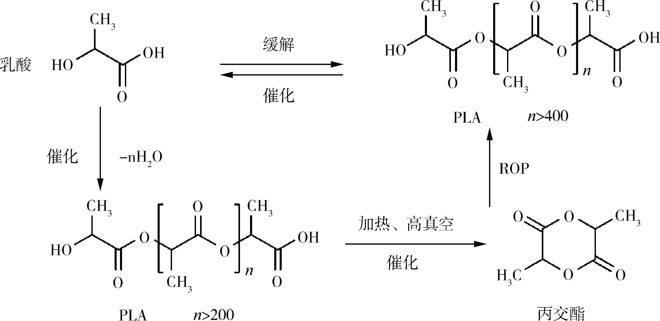
综述了聚乳酸(PLA)复合材料在骨组织工程材料中的研究进展;总结了国内外关于PLA复合材料的组成成分、合成工艺、加工方法及相对应的力学、生物学性能等方面的研究概况;梳理了PLA复合材料组成、结构与性能的相互关系,对PLA复合材料在骨组织修复材料中的应用发展前景进行了展望。
中图分类号:
韦宗辰, 郗悦玮, 翁云宣. 聚乳酸基复合骨组织修复材料的研究现状及进展[J]. 中国塑料, 2021, 35(9): 136-146.
WEI Zongchen, XI Yuewei, WENG Yunxuan. Research Progress in Poly(lactic acid)⁃based Composite Materials for Bone Tissue Engineering[J]. China Plastics, 2021, 35(9): 136-146.
| 材料分类 | 产品 | 优势 | 缺陷 |
|---|---|---|---|
| 金属基骨组织修复材料 | 主要有镍铬不锈钢、钴铬钼合金和钛及其合金,如骨钉、骨板、人工关节、人工血管和人工晶状体等。 | 耐蚀性和化学稳定性,力学上具有适宜的强度、韧性、耐磨性和耐疲劳性能,易加工成各种复杂形状,价格便宜和使用方便。 | 生物惰性材料不与组织相结合,既不能被吞噬系统所吞噬,也无法作为异物被排出体外。 |
| 无机非金属基骨组织修复材料 | 生物活性玻璃、生物陶瓷、可吸收缝合线等多种生物活性材料。 | 具备生物活性,即材料本身无毒,又具有高度的生物相容性,且在体内可促进缺损组织再生。 | 与金属基材料相比,无机非金属材料可塑性较差,力学性能不佳,成型性也较差。 |
| 高分子基骨组织修复材料 | 由PLA、橡胶、聚乙烯、聚丙烯等可降解和不可降解高分子制备的人工心脏瓣膜、人工骨等。 | 分子键较强,具有较高的化学稳定性、力学强度、可加工性和耐磨损性能。 | 大部分高分子基材料没有生物活性,部分高分子可能引起炎症及免疫反应。 |
| 有机/无机复合骨组织修复材料 | 组织工程支架材料、原位组织再生材料、可降解复合细胞和(或)生长因子材料等。 | 有机材料与无机材料相结合,既具备生物活性,又具备较好的力学性能。 | 制备难度高,相比其他的材料运用范围较小,针对性高,还需进一步研究发展。 |


| 1 | HO⁃SHUI⁃LING A, BOLANDER J, RUSTOM L E. et al. Bone Regeneration Strategies: Engineered Scaffolds, Bioactive Molecules and Stem Cells Current Stage and Future perspectives[J]. Biomaterials, 2018,180: 143⁃162. |
| 2 | GOMEZ⁃BARRENA E, ROSSET P, LOZANO D, et al. Bone Fracture Healing: Cell Therapy in Delayed Unions and Nonunions[J]. Bone, 2015,70: 93⁃101. |
| 3 | CANCEDDA R, GIANNONI P, MASTROGIACOMO M. A Tissue Engineering Approach to Bone Repair in Large Animal Models and in Clinical Practice. Biomaterials[J], 2007,28(29): 4 240⁃4 250. |
| 4 | NARAYANAN G, VERNEKAR V N, KUYINU E L, et al. Poly (lactic acid)⁃based Biomaterials for Orthopaedic Regenerative Engineering[J]. Advanced Drug Delivery Review, 2016, 107: 247⁃276. |
| 5 | MURUGAN R, RAMAKRISHNA S. Development of Nanocomposites for Bone Grafting[J]. Composites Science and Technology, 2005, 65(15/16): 2 385⁃2 406. |
| 6 | SALGADO A J, COUTINHO O P, REIS R L. Bone Tissue Engineering: State of the Art and Future Trends[J]. Macromol Biosci, 2004, 4(8): 743⁃765. |
| 7 | JOSE M V, THOMAS V, JOHNSON K T, et al. Aligned PLGA/HA Nanofibrous Nanocomposite Scaffolds For Bone Tissue Engineering[J]. Acta Biomater, 2009, 5(1): 305⁃315. |
| 8 | STAIGER M P, PIETAK A M, HUADMAI J, et al. Magnesium and Its Alloys as Orthopedic Biomaterials: A Rview[J]. Biomaterials, 2006, 27(9): 1 728⁃1 734. |
| 9 | NIINOMI M. Design and Development of Metallic Biomaterials with Biological and Mechanical Biocompatibility[J]. Journal of Biomedical Materials Research Part A, 2019,107(5): 944⁃954. |
| 10 | SANTOS E, ORIVEGORKA, JOSE LUIS PEDRAZ, et al. Cell⁃Biomaterial Interaction: Strategies to Mimic the Extracellular Matrix[J]. Biomimetics, 2011,6: 642⁃650. |
| 11 | SINGHVI M S, ZINJARDE S S, GOKHALE D V. Polylactic Acid: Synthesis and Biomedical Applications[J]. Journal of Applied Microbiology, 2019, 127(6): 1 612⁃1 626. |
| 12 | WANG J Z, YOU M L, DING Z Q, et al. A Review of Emerging Bone Tissue Engineering Via PEG Conjugated Biodegradable Amphiphilic Copolymers[J]. Mater Sci Eng C Mater Biol Appl, 2019,97: 1 021⁃1 035. |
| 13 | HOLLISTER S J, MURPHY W L. Scaffold Translation: Barriers between Concept and Clinic[J]. Tissue Engineering Part B: Reviews, 2011, 17(6): 459⁃474. |
| 14 | GRITSCH L, CONOSCENTI G, CARRUBBA L, et al. Polylactide⁃based Materials Science Strategies to Improve Tissue⁃material Interface without the use of Growth Factors or Other Biological Molecules[J]. Materials Science &Engineering C⁃Materials for Biological Applications, 2019,94: 1 083⁃1 101. |
| 15 | MIGUEZ⁃PACHECO V, HENCH L L, BOCCACCINI A R. Bioactive Glasses beyond Bone and Teeth: Emerging Applications in Contact with Soft Tissues[J]. Acta Biomater, 2015,13: 1⁃15. |
| 16 | BAINO F, NOVAJRA G, BOCCACCINI A R, et al. Bioactive Glasses: Special Applications Outside the Skeletal System[J]. Journal of Non⁃Crystalline Solids, 2016,432: 15⁃30. |
| 17 | XIAO Y F, YANG Y, LI J F, et al. Porous Composite Calcium Citrate/Polylactic Acid Materials with High Mineralization Activity and Biodegradability for Bone Repair Tissue Engineering[J]. International Journal of Polymeric Materials, 2021,70(7):507⁃520. |
| 18 | BHARADWAZ A,JAYASURIYA A C. Recent Trends in the Application of Widely Used Natural and Synthetic Polymer Nanocomposites in Bone Tissue Regeneration[J]. Mater Sci Eng C Mater Biol Appl, 2020,110: 110698. |
| 19 | CHEN J, ZHANG T, HUA W, et al. 3D Porous Poly(lactic acid)/Regenerated Cellulose Composite Scaffolds Based on Electrospun Nanofibers for Biomineralization[J].2019,585:124048 |
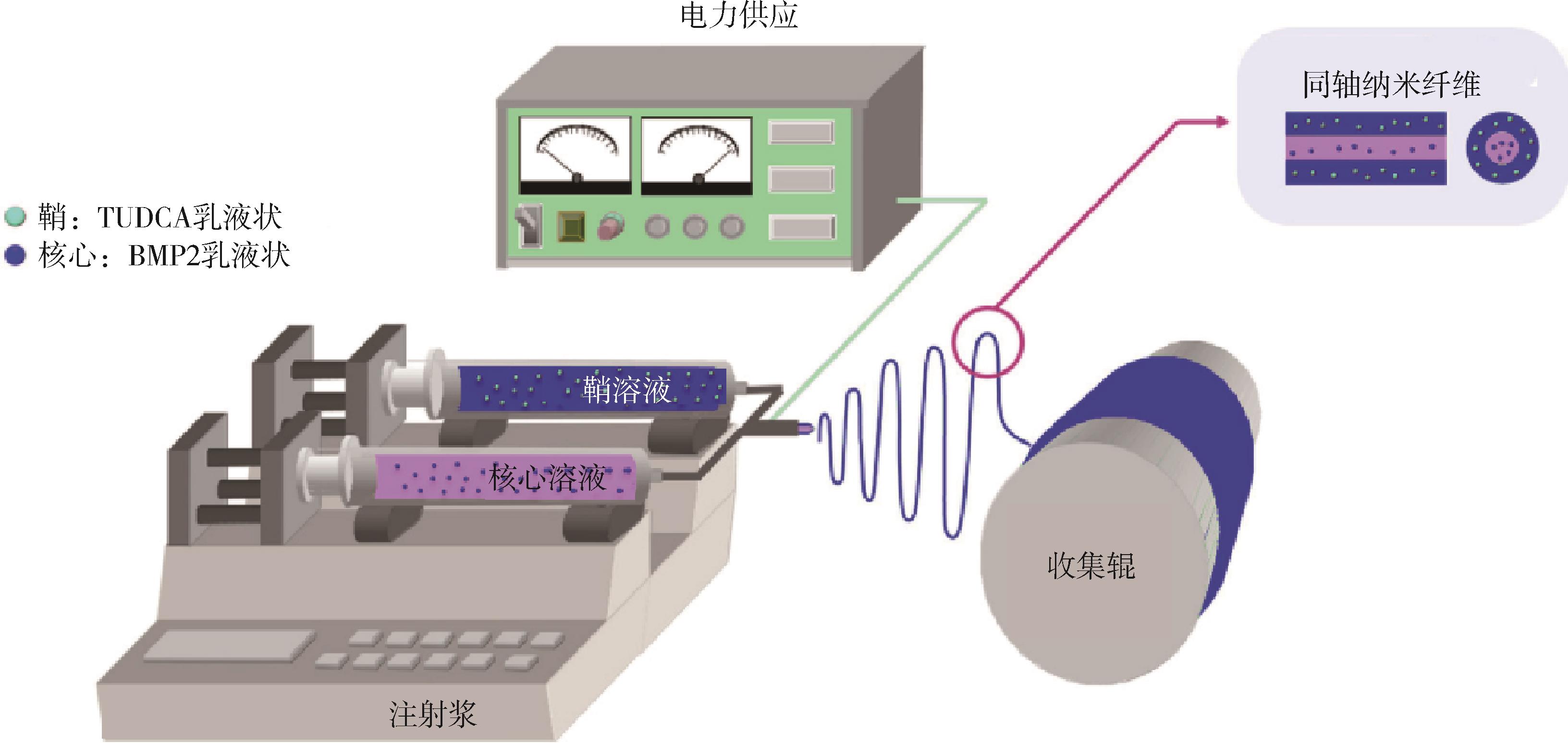
| 20 | DEVAL P B, MIN H K, HO P, et al. Coaxially Fabricated Polylactic Acid Electrospun Nanofibrous Scaffold for Sequential Release of Tauroursodeoxycholic Acid and Bone Morphogenic Protein2 to Stimulate Angiogenesis and Bone Regeneration[J]. Chemical Engineering Journal, 2020,389:123470. |
| 21 | DWEK J. The Periosteum: What Is It, Where Is It, And What Mimics It In Its Absence? [J]. Skeletal Radiol, 2010,39(4): 319⁃323. |
| 22 | SYED⁃PICARD F N, SHAH G A, COSTELLO B J, et al. Regeneration of Periosteum by Human Bone Marrow Stromal Cell Sheets[J]. Journal of Oral and Maxillofacial Surgery, 2014,72(6): 1 078⁃1 083. |
| 23 | WU L, GU Y, LIU L, et al. Hierarchical Micro/Nanofibrous Membranes of Sustained Releasing VEGF for Periosteal Regeneration[J]. Biomaterials, 2020,227: 119555. |
| 24 | COLNOT C, ZHANG X, KNOTHE T M. Current Insights on the Regenerative Potential of the Periosteum: Molecular, Cellular, and Endogenous Engineering Approaches[J]. Journal of Orthopaedic Research, 2012, 30(12): 1 869⁃1 878. |
| 25 | YANG Y Q, TAN Y Y, WONG R, et al. The Role of Vascular Endothelial Growth Factor in ossification[J]. Int J Oral Sci, 2012,4(2): 64⁃68. |
| 26 | HU K, YANG M, XU Y ,et al. Cell Cycle Arrest, Apoptosis, and Autophagy Induced by Chabamide in Human Leukemia Cells[J]. Chinese Herbal Medicines, 2016,8(1): 30⁃38. |
| 27 | LEE K,SILVA E A, MOONEY D J. Growth Factor Delivery⁃based Tissue Engineering: General Approaches and A Review of Recent Developments[J]. Journal of the Royal Society Interface, 2011. 8(55): 153⁃170. |
| 28 | MAES C, CARMELIET G, SCHIPANI E. Hypoxia⁃driven Pathways in Bone Development, Regeneration and Disease[J]. Nature Reviews Rheumatology, 2012, 8(6): 358⁃366. |
| 29 | YIN Z, CHEN X, CHEN J L, et al. The Regulation of Tendon Stem Cell Differentiation by the Alignment of Nanofibers[J]. Biomaterials, 2010,31(8): 2 163⁃2 175. |
| 30 | RUSSO V, ANCORA M, WYRWA R, et al. Tendon Biomimetic Electrospun PLGA Fleeces Induce an Early Epithelial⁃Mesenchymal Transition and Tenogenic Differentiation on Amniotic Epithelial Stem Cells[J]. Cells, 2020,9(2):303. |
| 31 | ZEUGOLIS D I, CHAN J C Y, PANDIT A. Tendons: Engineering of Functional Tissues[M].Heidelberg:Springer⁃Verlag,in Tissue Engineering,2011: 537⁃572. |
| 32 | LONGO U G, LAMBERTI A, MAFFULLI N, et al. Tendon Augmentation Grafts: A Systematic Review[J]. British Medical Bulletin, 2010,94: 165⁃188. |
| 33 | DENG D, WANG W, WANG B, et al. Repair of Achilles Tendon Defect With Autologous ASCs Engineered Tendon in A Rabbit Model[J]. Biomaterials, 2014,35(31): 8 801⁃8 809. |
| 34 | WANG F, ZHANG B, ZHOU L, et al. Imaging Dendrimer⁃Grafted Graphene Oxide Mediated Anti⁃miR⁃21 Delivery With an Activatable Luciferase Reporter[J]. ACS Applied Materials & Interfaces, 2016,8(14): 9 014⁃9 021. |
| 35 | WALDEN G, LIAO X, DONELL S, et al. A Clinical, Biological, and Biomaterials Perspective into Tendon Injuries and Regeneration[J]. Tissue Engineering Part B⁃Reviews, 2017,23(1): 44⁃58. |
| 36 | AMIRSADEGHI A, JAFARI A, EGGERMONT L J, et al. Vascularization Strategies for Skin Tissue Engineering[J]. Biomaterials Science, 2020,8(15): 4 073⁃4 094. |
| 37 | ZHANG X, BOGDANOWICZ D,ERISKEN C, et al. Biomimetic Scaffold Design for Functional and Integrative Tendon Repair[J]. Journal of Shoulder Elbow Surgery/American Shoulder and Elbow Surgeons, 2012,21(2):266⁃277. |
| 38 | ZHANG C, YUAN H, LIU H, et al. Well⁃aligned Chitosan⁃based Ultrafine Fibers Committed Teno⁃lineage Differentiation of Human Induced Pluripotent Stem Cells for Achilles Tendon Regeneration[J]. Biomaterials, 2015,53: 716⁃730. |
| 39 | THAYER P S, VERBRIDGE S S, DAHLGREN L A, et al. Fiber/Collagen Composites for Ligament Tissue Engineering: Influence of Elastic Moduli of Sparse Aligned Fibers on Mesenchymal Stem Cells[J]. Journal of Biomedical Materials Research Part A, 2016,104(8): 1 894⁃1 901. |
| 40 | DEEPTHI S, NIVEDHITHA SUNDARAM M, DEEPTI KADAVAN J, et al. Layered Chitosan⁃collagen Hydrogel/Aligned PLLA Nanofiber Construct for Flexor Tendon Regeneration[J]. Carbohydrate Polymers:Scientific and Technological Aspects of Industrially Important Polysaccharides, 2016,153: 492⁃500. |
| 41 | TAMAYOL A, AKBARI M, ANNABI N, et al. Fiber⁃based Tissue Engineering: Progress, Challenges, and Opportunities[J]. Biotechnology Advances, 2013, 31(5): 669⁃687. |
| 42 | MCCULLEN S D, HASLAUER C M, LOBOA E G. Fiber⁃reinforced scaffolds for Tissue Engineering and Regenerative Medicine: Use of Traditional Textile Substrates to Nanofibrous arrays[J]. Journal of Materials Chemistry, 2010,20(40):8 776⁃8 788. |
| 43 | VAISHYA R, PATRALEKH M K, BIJUKCHHE A R, et al. The Top 10 Arthroplasty Articles Published in Last 10 Years by Indian Authors[J]. Journal of Clinical Orthopaedics and Trauma, 2018,9(1): 94⁃100. |
| 44 | RIBEIRO V P, SILVA⁃CORREIA J, NASCIMENTO A I, et al. Silk⁃based Anisotropical 3D Biotextiles for Bone Regeneration[J]. Biomaterials, 2017,123: 92⁃106. |
| 45 | WU S H, ZHOU R, ZHOU F, et al. Electrospun Thymosin Beta⁃4 loaded PLGA/PLA nanofiber/ microfiber Hybrid Yarns for Tendon Tissue Engineering Application[J]. Materials Science & Engineering, 2020,106(1):110268. |
| 46 | HE Y, XU W H, ZHANG H, et al. Constructing Bone⁃Mimicking High⁃Performance Structured Poly(lactic acid) by an Elongational Flow Field and Facile Annealing Process[J]. ACS Applied Materials & Interfaces, 2020,12(11): 13 411⁃13 420. |
| 47 | BANKOLE I O, ZAHEDI S A, ADEOYE A O M, et al. 3D Printing of Bone Scaffolds with Hybrid Biomaterials[J].Composites Part B, 2019,158:428⁃436 |
| 48 | WU D, SPANOU A, DIEZ⁃ESCUDERO A, et al. 3D⁃printed PLA/HA Composite Structures as Synthetic Trabecular Bone: A Feasibility Study Using Fused Deposition Modeling[J]. Journal of the Mechanical Behavior of Biomedical Materials, 2020,103: 103608. |
| 49 | ALAMFAHAD, VARADARAJAN K M, KUMAR S, et al. 3D Printed Polylactic Acid Nanocomposite Scaffolds for Tissue Engineering Applications[J].Materials Science & Engineering c⁃materials for Biological Applications, 2020,81:106203 |
| 50 | SILVA S S, MANO J F, REIS R L. Potential Applications of Natural Origin Polymer⁃based Systems in Soft tissue Regeneration[J]. Critical Reviews in Biotechnology, 2010,30(3): 200⁃221. |
| 51 | YOUNES I, RINAUDO M. Chitin and Chitosan Preparation From Marine Sources. Structure, Properties and Applications[J]. Marine Drugs, 2015,13(3): 1 133⁃1 174. |
| 52 | NISHINO R, KAKIZAKI H, FUKUSHIMA H, et al. Distribution of Chitinolytic Enzymes in the Organs and cDNA Cloning of Chitinase Isozymes from the Liver of Golden Cuttlefish Sepia esculenta[J]. Advances in Bioscience and Biotechnology, 2017,8(10): 361⁃377. |
| 53 | ZHOU D, ZHANG L, GUO S. Mechanisms of Lead Biosorption on Cellulose/Chitin Beads[J]. Water Research, 2005, 39(16): 3 755⁃3 762. |
| 54 | MUXIKA A, ETXABIDE A, URANGA J, et al. Chitosan as A Bioactive Polymer: Processing, Properties and Applications[J]. International Journal of Biological Macromolecules, 2017,105(2):1 358⁃1 368. |
| 55 | BHATNAGAR A, SILLANPAA M. Applications of Chitin⁃ and chitosan⁃derivatives for the Detoxification of Water and Wastewater⁃a Short Review[J]. Advances in Colloid & Interface Science, 2009,152(1/2): 26⁃38. |
| 56 | BRIGHAM C J. Chitin and Chitosan: Sustainable, Medically Relevant Biomaterials[J]International Journal of Biotechnology for Wellness Industries, 2017,6:41⁃47. |
| 57 | KURITA K. Chitin and Chitosan: Functional Biopolymers from Marine Crustaceans[J]. Mar Biotechnol (NY), 2006, 8(3): 203⁃226. |
| 58 | FOSTER L J, HO S, HOOK J, et al. Chitosan as A Biomaterial: Influence of Degree of Deacetylation on Its Physiochemical, Material and Biological Properties[J]. Public Library of Science One, 2015,10(8): 135⁃153. |
| 59 | YUSOF N L B M, YUSOF M, WEE A, et al. Flexible Chitin Films as Potential Wound⁃dressing Materials: Wound Model Studies[J]. Chemistry, 2003,66 A(2):224⁃232. |
| 60 | RASAL R M, JANORKAR A V, HIRT D E. Poly(lactic acid) Modifications[J]. Progress in Polymer Science, 2010,35(3): 338⁃356. |
| 61 | KEHAIL A A, BRIGHAM C J. Anti⁃biofilm Activity of Solvent⁃Cast and Electrospun Polyhydroxyalkanoate Membranes Treated with Lysozyme[J]. Journal of Polymers and the Environment, 2017,26(1): 66⁃72. |
| 62 | FREED L E, BORENSTEIN J T, MOUTOS F T, et al. Advanced material Strategies for Tissue Engineering Scaffolds[J]. Adv Mater, 2009, 21(32/33):3 410⁃3 418. |
| 63 | RAZAK S I A, SHARIF N F A, RAHMAN W A W A. Biodegradable Polymers and Their Bone Applications: A Review[J]. International Journal of Basic & Applied Sciences.2012,12(1):31⁃49. |
| 64 | BIOLOGY D O, DIVISION H S T, DIVISION E S. Applications of Polyhydroxyalkanoates in the Medical Industry[J]. International Journal of Biotechnology for Wellness Industries, 2012,1:53⁃60. |
| 65 | MUNIRAH S, KIM S H, RUSZYMAH B H, et al. The Use of Fibrin and Poly(lactic⁃co⁃glycolic acid) Hybrid Scaffold for Articular Cartilage Tissue Engineering: an in Vivo Analysis[J]. European Cells & Materials, 2008,15: 41⁃52. |
| 66 | CARRASCO F, SANTANA O O, MASPOCH M L, et al. Processing of Poly(lactic acid): Characterization of Chemical Structure, Thermal Stability and Mechanical Properties[J]. Polymer Degradation and Stability, 2010,95(2): 116⁃125. |
| 67 | BALAKRISHNAN H, HASSAN A, WAHIT M, et al. Novel Toughened Polylactic Acid Nanocomposite: Mechanical, Thermal and Morphological Properties[J]. Materials & Design, 2010,31(7): 3 289⁃3 298. |
| 68 | WAHL D A, CZERNUSZKA J T. Collagen⁃hydroxyapatite Composites for Hard Tissue Repair[J]. Eur Cell Mater, 2006,11: 43⁃56. |
| 69 | SERRA T, PLANELL J A, NAVARRO M, et al. 3D Printed PLA⁃based Scaffolds: A Versatile Tool in Regenerative Medicine[J]. Organogenesis, 2013,9(4): 239⁃244. |
| 70 | GREGOR A, FILOVA E, NOVAK M, et al. Designing of PLA Scaffolds for Bone Tissue Replacement Fabricated by Ordinary Commercial 3D Printer[J]. Journal of Biological Engineering, 2017,11: 31. |
| 71 | DOROZHKIN S V. Calcium Orthophosphate⁃Based Bioceramics[J]. Materials (Basel), 2013. 6(9): 3 840⁃3 942. |
| 72 | TRACY B M, DOREMUS R H. Direct Electron Microscopy Studies of The Bone⁃hydroxylapatite Interface[J]. Journal of Biomedical Materials Research,1984,18(7):719⁃726. |
| 73 | HONG Z, ZHANG P, HE C, et al. Nano⁃composite of Poly(L⁃lactide) and Surface Grafted Hydroxyapatite: Mechanical Properties and Biocompatibility[J]. Biomaterials, 2005,26(32): 6 296⁃6 304. |
| 74 | WANG H, LI Y, ZUO Y, et al. Biocompatibility and Osteogenesis of Biomimetic Nano⁃hydroxyapatite/Polyamide Composite Scaffolds for Bone Tissue Engineering[J]. Biomaterials, 2007,28(22): 3 338⁃3 348. |
| 75 | STIPNIECE L, RJABOVS V, JUHNEVICA I, et al. Development of Functionalized Hydroxyapatite/poly(vinyl alcohol) Composites[J]. Journal of Crystal Growth, 2016,444: 14⁃20. |
| 76 | HABRAKEN W, HABIBOVIC P, EPPLE M, et al. Calcium Phosphates in Biomedical Applications: Materials for The Future[J].Materials Today, 2016,19(2): 69⁃87. |
| 77 | MYGIND T, STIEHLER M, LI H, et al. Mesenchymal Stem Cell Ingrowth and Differentiation on Coralline Hydroxyapatite Scaffolds[J]. Biomaterials, 2007,28(6): 1 036⁃1 047. |
| 78 | YOSHIKAWA H, MYOUI A. Bone Tissue Engineering with Porous Hydroxyapatite Ceramics[J]. Journal of Artificial Organs, 2005,8(3): 131⁃136. |
| 79 | SUN F, ZHOU H, LEE J. Various Preparation Methods of Highly Porous Hydroxyapatite/Polymer Nanoscale Biocomposites for Bone Regeneration[J]. Acta Biomater, 2011,7(11): 3 813⁃3 828. |
| 80 | CHAKRAVARTY J, RABBI M F, CHALIVENDRA V, et al. Mechanical and Biological Properties of Chitin/Polylactide (PLA)/Hydroxyapatite (HAP) Composites Cast Using Ionic Liquid Solutions[J]. International Journal of Biological Macromolecules, 2020,151: 1 213⁃1 223. |
| [1] | 于昌永, 辛忠. 基于六氢邻苯二甲酸盐的α/β复合成核剂对聚丙烯性能的影响[J]. 中国塑料, 2022, 36(7): 121-128. |
| [2] | 冯冰涛, 王晓珂, 张信, 孙国华, 汪殿龙, 侯连龙, 马劲松. 连续碳纤维增强热塑性复合材料制备与应用研究进展[J]. 中国塑料, 2022, 36(7): 165-173. |
| [3] | 谭立钦, 刘伟区, 梁利岩, 王硕, 冯志强, 林家明. 含巯基聚硅氧烷改性环氧树脂的制备及性能[J]. 中国塑料, 2022, 36(7): 21-29. |
| [4] | 宋银宝, 杨建军, 李传敏. PDMS/SiC功能梯度复合材料性能与制造精度研究[J]. 中国塑料, 2022, 36(7): 30-36. |
| [5] | 徐杰, 钟进福, 童晓茜, 李广富, 付栋梁, 李城城. 端羧基修饰单宁酸/没食子酸环氧树脂复合材料的制备与性能研究[J]. 中国塑料, 2022, 36(7): 44-50. |
| [6] | 沈雪梅, 朱小龙, 胡燕超, 宋任远, 张现峰, 李席. 静电喷雾法制备聚乳酸/布洛芬微球及其性能研究[J]. 中国塑料, 2022, 36(7): 61-67. |
| [7] | 周舒毅, 朱敏, 刘忆颖, 曹舒惠, 蔡启轩, 聂慧, 张玉霞, 周洪福. 高分子止血材料研究进展[J]. 中国塑料, 2022, 36(7): 74-84. |
| [8] | 李凯泽, 辛勇. 改性碳纳米管增强热塑性聚氨酯复合材料的性能研究[J]. 中国塑料, 2022, 36(6): 1-5. |
| [9] | 邵琳颖, 郗悦玮, 翁云宣. 可降解聚乳酸复合材料研究进展[J]. 中国塑料, 2022, 36(6): 155-164. |
| [10] | 杨小龙, 陈文静, 李永青, 闫晓堃, 王修磊, 谢鹏程, 马秀清. 导电型聚合物/石墨烯复合材料的研究进展[J]. 中国塑料, 2022, 36(6): 165-173. |
| [11] | 王帅, 张玉迪, 杨富凯, 徐新宇. 聚酰亚胺/多壁碳纳米管泡沫材料的制备及性能研究[J]. 中国塑料, 2022, 36(6): 39-45. |
| [12] | 王金业, 唐博虎, 杨立宁, 谢猛, 郭泽朝, 杨光. PA12试件多射流熔融成型工艺研究[J]. 中国塑料, 2022, 36(6): 81-86. |
| [13] | 孙文博, 信春玲, 何亚东, 翟玉娇, 闫宝瑞. 玻璃纤维增强PBT微发泡工艺对其制品泡孔结构的影响[J]. 中国塑料, 2022, 36(5): 1-7. |
| [14] | 王镕琛, 张恒, 孙焕惟, 段书霞, 秦子轩, 李晗, 朱斐超, 张一风. 医疗卫生用聚乳酸非织造材料的制备及其亲水改性研究进展[J]. 中国塑料, 2022, 36(5): 158-166. |
| [15] | 王轲, 龙春光. PE⁃UHMW/海泡石纤维复合材料的力学性能与摩擦学性能研究[J]. 中国塑料, 2022, 36(5): 19-23. |
| 阅读次数 | ||||||
|
全文 |
|
|||||
|
摘要 |
|
|||||
京公网安备11010802034965号
京ICP备13020181号-2